Fruit Fly
Neural Canvas Puts 166,700 Fruit-Fly Neurons in Your Browser
Xenova’s Neural Canvas turns the new MaleCNS wiring map into an interactive browser experiment while separating connectivity, simulated dynamics, and crafted motion.
 Akmal Alif · 10 September 2026 MYT
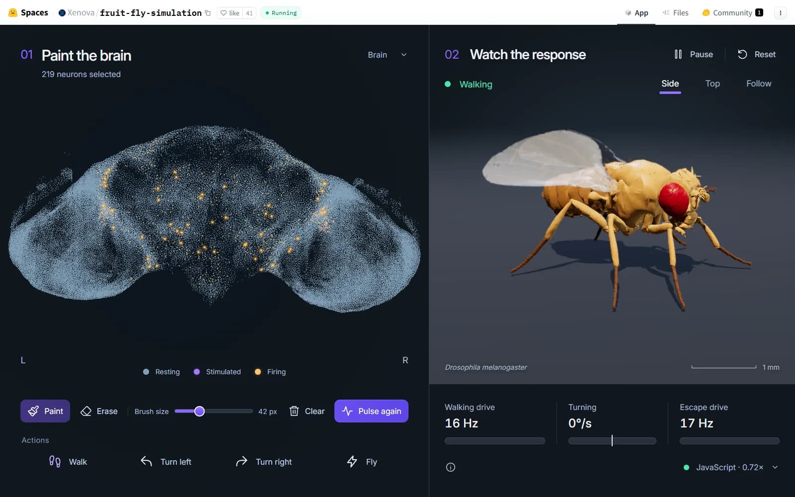
At first glance, Neural Canvas feels almost impossible: a browser tab fills with a point-cloud brain, you paint a patch of neurons, and an articulated fruit fly starts to move. The important story is not that a fly has been uploaded to the web. It is that Xenova has turned a new, unusually complete wiring dataset into a transparent experiment that runs on a reader’s own machine.
The link that has been circulating under 0xSojalSec’s account is a duplicate of Xenova’s canonical Space. As of September 10, 2026, Hugging Face reports the same repository commit for both Spaces. Credit, source inspection and future updates should therefore point to Xenova’s original.
A map of connections, not a captured fly
The underlying resource is MaleCNS, the newly released connectome of an adult male Drosophila melanogaster. The Google Research release describes a proofread map spanning the brain and ventral nerve cord, with more than 166,000 neurons and roughly 125 million synaptic contacts. The accompanying Cell paper reports 166,691 neurons and 11,691 cell types.
Xenova’s packaged model retains 166,700 MaleCNS records, 124,177,617 synapses and 25,582,938 directed edges. Those last two numbers describe different levels of the graph: many synaptic contacts can be aggregated into one directed connection between a source and a target. The demo preserves the large-scale wiring resource while packaging it for interactive computation.
A connectome is structural evidence. It records which reconstructed neurons contact which others and with what count; it does not preserve every membrane property, chemical state, learned parameter, sensory history or ongoing electrical event from a living animal. Neural Canvas is therefore a connectome-based simulation, not a living fly, a mind upload or a claim that a biological brain has been captured.
How 166,700 records run in one browser tab
The canonical README lays out a deliberately local architecture. After the reader chooses “Download & start,” about 76 MB of weights and anatomy are loaded and cached. No API key or remote model service is required. WebGPU kernels handle the fast path when supported; a JavaScript reference implementation provides a fallback. Three.js and WebGL2 render the neural point cloud and the fly.
1. Connectivity becomes a sparse computational graph
The connection data is arranged so spikes can propagate through outgoing edges without materialising a dense 166,700-by-166,700 matrix. The WebGPU path uses sparse queues and fused update kernels, while the JavaScript path implements the same reference calculation. That engineering choice is what makes an otherwise unwieldy graph usable in an ordinary web interface.
2. A spiking model supplies the dynamics
Static wiring cannot animate itself. Neural Canvas adds a leaky integrate-and-fire model adapted from earlier fruit-fly simulation research. Each step integrates membrane state, delayed synaptic input, thresholds, resets and refractory periods. Neurotransmitter labels supply simplified excitatory or inhibitory signs. The model is deterministic at its documented seed, but it still contains chosen parameters and omissions: receptor-specific dynamics, gap junctions, intrinsic cell diversity and much neuromodulation are not represented.
3. Firing rates drive a crafted body controller
The visible fly is not a biomechanical consequence calculated directly from every spike. The application gathers activity from selected motor-related populations, converts firing rates into six control signals, and maps those signals to walking speed, yaw and escape flight. Authored tripod foot trajectories, joint-limited inverse kinematics and a bounded flight controller then animate a measured skeleton.
What painting and the action buttons really do
Readers can paint neurons directly or use presets labelled Walk, Turn left, Turn right and Fly. The presets are engineered interventions: Walk stimulates bilateral LC9 neurons, turns stimulate LC9 plus same-side DNa02, and Fly stimulates bilateral LC4. The activity still propagates through the simulated network, but the target cells and pulse envelopes were selected by the developer. They are not natural sensory scenes delivered through a complete virtual eye.
This distinction matters when interpreting the animation. A turn can remain visible even when recurrent transmission is disabled because DNa02 is stimulated directly. The movement is useful feedback about the chosen readout pipeline; it is not, by itself, evidence that the connectome predicts an animal’s behaviour.
The honest boundary around the moving fly
Motion is authored and open-loop. The demo has no individual muscles, ground-contact physics or aerodynamic simulation.
The body does not send touch, proprioception or visual consequences back into the neural network, so action cannot alter the next sensory input.
The transferred spiking dynamics have not been validated for the MaleCNS connectome. Validation of the earlier reference model does not validate this new transfer.
The visual body is a female NeuroMechFly specimen, while the wiring map is male. It is a rendering asset, not a sex-matched digital organism.
These are not footnotes to hide. They define what the experiment is. Neural Canvas shows recorded connectivity influencing simulated spikes, then uses selected rates to drive an illustrative controller. It does not model a closed perception–action loop, and its movements are not validated predictions of fly behaviour.
The interface helps readers keep those layers visible. The left panel reports which neurons are selected and colours resting, stimulated and firing points separately. The right panel labels the fly’s state and exposes walking, turning and escape-drive rates alongside the active compute path. Those readouts make the causal chain easier to inspect, but they remain instrumentation from the model. A glowing neuron means the implemented equations crossed a threshold; a walking label means the controller crossed a chosen activation level. Neither display substitutes for measurements from an animal.
Why the experiment still matters
The demo compresses several difficult systems into something inspectable: a public connectome, sparse computation, browser-local acceleration, a neural visualisation and an articulated body. Because it exposes the wiring source, parameters, action targets, licenses and limitations, it gives researchers and programmers a concrete baseline they can criticise and extend instead of a cinematic claim they can only watch.
That makes Neural Canvas valuable as an instrument for asking better questions. How sensitive are firing patterns to synaptic signs and time constants? Which activity survives when wiring is randomised? Can observed neural recordings calibrate the dynamics? What changes when a body sends sensory consequences back into the model? Each question can become a controlled experiment.
What should come next
Compare simulated activity with recorded responses for identified cells and stimuli, using held-out measurements rather than visual plausibility.
Publish sensitivity analyses across seeds, thresholds, delays, transmitter assumptions and connection perturbations.
Separate the connectome’s contribution from the controller by testing random wiring, degree-preserving rewires and readout-only baselines.
Add a closed loop in which body motion changes sensory input, then measure stability and behaviour under controlled environments.
Neural Canvas does not settle whether connectome structure is enough for useful computation. It gives the community a legible place to test that proposition. A separate EXEPERT field report examines how Eris pushed the same newly available connectome toward next-token prediction—a bolder claim with a very different evidence boundary.